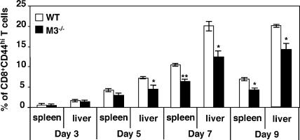
Figure 3.
Decreased frequency of effector CD8+ T cells in M3−/− mice during LM infection. Splenocytes and hepatic leukocytes were harvested from LM-infected M3−/− and M3+ mice (N10) on the indicated days and stained with mAbs specific for CD8α, CD4, and CD44. Percentages of CD44high CD8+ T cells were determined by flow cytometric analysis. Data shown are the mean ± SEM of three to eight animals per interval.

