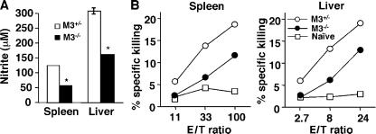
Figure 5.
Defective nitrite production and NK cell activity in M3−/− mice during the early phase of LM infection. M3−/− and M3+/− littermate controls (N6) were infected with 5 × 10³ CFU of LM. (A) Measurement of nitrite production. 3 d after infection, splenocytes and hepatic leukocytes were isolated and stimulated with HKLM. After 48 h of incubation, the concentration of nitrite as a surrogate marker for the production of reactive nitrogen was determined with Greiss reagent. Bars represent mean ± SEM of two mice per group and are representative of four independent experiments. (B) NK cell cytotoxicity assay. Splenocytes and hepatic lymphocytes were isolated from day 3 LM-infected mice and used as effectors. Varying numbers of effectors were combined with a constant number of 51Cr-labeled YAC-1 cells in a standard 4-h cytotoxicity assay. Each data line represents the mean from two mice and is representative of four independent experiments.

