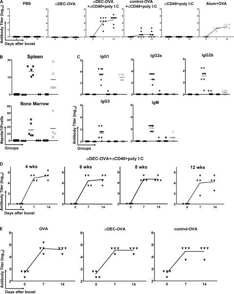
Figure 2.
Immunization with anti–DEC-OVA fusion mAb primes helper T cells for antibody responses. (A) Anti-NP IgG antibody titers before and 7 or 14 d after an i.p. boost injection with 1 μg OVA-NP11 in PBS. Immunization conditions are displayed at the top. Symbols represent individual mice, and the curves represent the mean value of each group. (B) Antibody-secreting cells in the spleen or bone marrow measured by ELISPOT on NP2-BSA 14 d after an i.p. boost with 1 μg OVA-NP11 in PBS. Symbols indicate individual mice. Initial immunization conditions were as follows: PBS (▪), anti–DEC-OVA (▴), anti–DEC-OVA plus anti-CD40 plus poly I:C (▾), control-OVA plus anti-CD40 plus poly I:C (♦), anti-CD40 plus poly I:C (•), and alum plus OVA (×). (C) Anti-NP antibody titers for individual IgG isotypes and IgM 14 d after an i.p. boost with 1 μg OVA-NP11 in PBS. The horizontal lines represent the mean value of each group. Symbols indicate individual mice and initial immunization conditions as described in B. (D) Anti-NP IgG antibody responses measured by ELISA in mice immunized with anti–DEC-OVA plus anti-CD40 plus poly I:C 4, 6, 8, or 12 wk before i.p. boosting with 1 μg OVA-NP11 in PBS. Antibody titers were measured as described in A. (E) Anti-OVA IgG antibody titers in mice immunized with anti–DEC-OVA plus anti-CD40 plus poly I:C and then boosted 6 wk later with 1 μg of either OVA, anti–DEC-OVA, or control-OVA. Antibody titers were measured as described in A.

