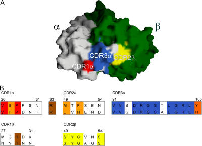
Figure 5.
Conserved residues from human and mouse NKT TCR CDR1α, CDR3α, and CDR2β loops form a contiguous conserved surface that is adjacent to the putative Ag-binding cavity. The different conserved CDR regions are colored to correspond to the sequence alignment of mouse and human CDR loops. All other conserved and semi-conserved residues are brown and orange, respectively, in the alignment and are not shown in the structure. The TcR α-chain is white and the TCR β-chain is green.

