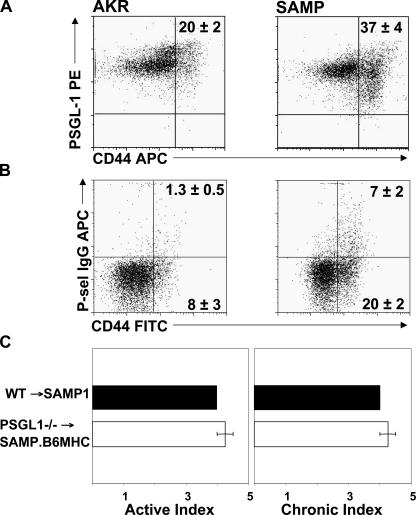
Figure 3.
A higher percentage of SAMP1/YitFc (SAMP) CD4+/CD44high MLN cells bind P-selectin–IgG compared with cells from uninflamed AKR control mice (AKR). (A) PSGL-1 expression and (B) P-sel–IgG binding to MLN cells were analyzed by flow cytometry and gated on forward light scatter (FSC), side light scatter (SSC), and CD4. Representative data obtained from four and three mice, respectively. (C) The severity of ileitis was determined in SAMP1.B6MHC mice reconstituted with PSGL-1–sufficient (WT) or deficient (PSGL−/−) bone marrow. Error bars show the mean ± SEM.

