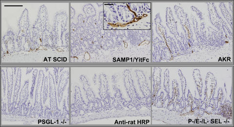
Figure 5.
Small intestinal endothelial cells express PSGL-1. Immunohistochemistry was performed on frozen terminal ileal sections from the indicated mouse strains as per Materials and methods. PSGL-1 expression was detected on leukocytes and in venules within the LP and submucosa of adoptively transferred SCID mice (AT SCID), SAMP1/YitFc, and AKR mice. Minimal or no expression was observed in PSGL-1–deficient (−/−) mice or in AKR ileum reacted with anti–rat HRP after isotype antibody injection (Anti–rat HRP), whereas robust expression was seen in P-, E-, L-selectin triple-deficient (−/−) mice, which lack rolling leukocytes. Bar, 200 μm. Inset, high power of SAMP1/YitFc submucosal venules. Bar, 20 μm.

