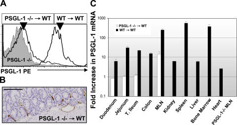
Figure 7.
Endothelial PSGL-1 expression localized to the small intestine and MLN of PSGL1−/− → WT bone marrow chimeric mice. (A) Peripheral blood leukocytes from PSGL-1–deficient or indicated bone marrow chimeric mice were incubated with PE anti–PSGL-1 (2PH1) antibody as per Materials and methods. Cells were gated on FSC, SSC, and CD3. (B) Endothelial PSGL-1 was detected by immunohistochemistry in mice reconstituted with PSGL-1–deficient bone marrow using mAb 4RA10. Bar, 150 μm. (C) PSGL-1 mRNA levels from indicated organs of bone marrow chimeric and PSGL1−/− mice. Indicated tissues were harvested, RNA was obtained, and real-time RT-PCR was performed as per Materials and methods. Representative histograms, tissues, and mRNA expression were obtained from at least four bone marrow chimeric mice per group and run in triplicate. Error bars show the mean ± SEM.

