Figure 9.
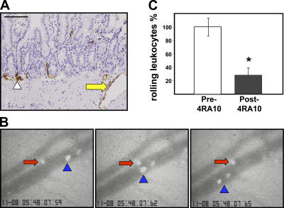
PSGL-1 blockade decreased splenocyte–endothelial interactions in the small intestinal vasculature. (A) Endothelial PSGL-1 signal was observed in small (arrowhead) and large vessels (yellow arrow) within the small intestinal muscularis and serosa. The larger serosal vessels were monitored via intravital microscopy. Bar, 200 μm. (B) Rolling CFSE-labeled PSGL-1–deficient cells (red arrows) proceed much slower than cells not interacting with wild-type vessels (blue arrowhead) before (B) PSGL-1 blockade. (C) A significant decrease in the number of rolling cells was observed after injection of 4RA10 (percent of normalized total rolling cells [error bars show the mean ± SD] before and after 4RA10 injection from four experiments in which one to three vessels/mouse were monitored; *, P < 0.05).

