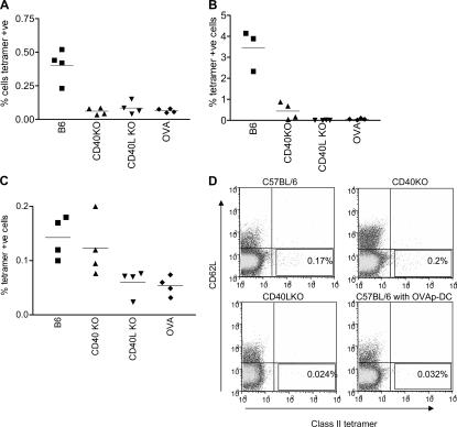
Figure 3.
CD40–CD40L signals are required for T cell priming. (A) C57BL/6, CD40KO, and CD154KO mice were immunized with H19-Env in CFA. 9 d later, spleens and draining LNs were taken and cells were stained with class II tetramers. Data from draining LNs are shown with each point representing the percentage of CD4 cells that were tetramer+ in an individual mouse, and the mean of the group is shown. (B) Splenocytes from immunized mice were activated with 1 μg/ml H19-Env peptide. After 4 d of culture, the percentage of tetramer+ cells in the CD4+ population was measured by FACS. (C) WT DC primed T cells in CD40KO mice. C57BL/6, CD40KO, and CD154KO mice were immunized with H19-Env peptide–pulsed, LPS-matured DCs from WT mice. The percentage of tetramer+ cells in the CD4+ population was measured by FACS in the draining LNs 6 d later. (A–C) Each point represents one mouse, and the line shows the mean of each group. Results are representative of three independent experiments. (D) Representative FACS dot plots from the experiment shown in C. Control mice were immunized with OVA-CFA (A and B) or OVA-pulsed DCs (C and D) and stained with the H19-Env tetramers.

