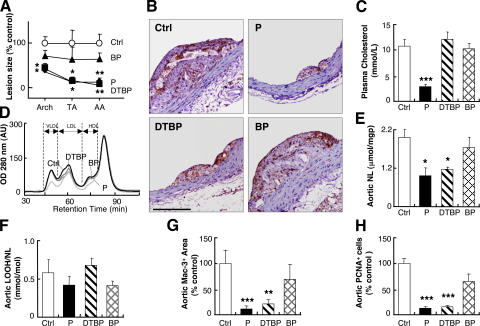
Figure 1.
Probucol and probucol dithiobisphenol, but not probucol bisphenol, inhibit atherosclerosis in Apoe−/− mice. (A) Lesion area in animals treated with probucol (P, ♦), probucol dithiobisphenol (DTBP, ▪), or probucol bisphenol (BP, ▴) compared with control (Ctrl, ○). n = 10 for each site, each using two, six, and three serial sections for arch, thoracic (TA), and abdominal aorta (AA), respectively. (B) Representative cross sections of abdominal aorta from control and the three treatment groups stained for macrophages indicating respective lesion size. Bar, 25 μm. (C) Plasma cholesterol, n = 10. (D) FPLC chromatograms of lipoproteins from pooled plasma (n = 10) of control, probucol, probucol dithiobisphenol, and probucol bisphenol animals. (E) Neutral lipids (NL, comprised of cholesterylesters and triglycerides) and (F) their hydroperoxides (LOOH) standardized to NL. n = 3 pools of five aortas per pool. (G) Average lesion area covered by Mac-3+ cells (i.e., macrophages) and (H) PCNA+ (i.e., proliferating) cells in the arch, thoracic, and abdominal aorta as compared with corresponding control. n = 3 for each site (three serial sections/site). *, P < 0.05; **, P < 0.01; ***, P < 0.001 compared with control.

