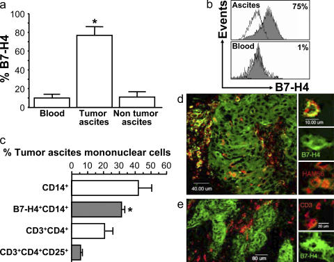
Figure 2.
Tumor-associated macrophages express B7-H4. (a and b) Tumor ascites macrophages express B7-H4. FACS analysis showed that ascites macrophages, but not control nonmalignant ascites macrophages, or fresh normal blood monocytes expressed B7-H4. Results are expressed as the mean of the percentage of B7-H4+ cells ± SEM in total macrophages. Filled histogram, B7-H4 staining; open histogram, isotype control. (c) High prevalence of B7-H4+ tumor macrophages in tumor ascites. FACS analysis was used to determine the prevalence of B7-H4+ tumor macrophages and CD3+CD4+CD25+ cells in tumor ascites (*, P < 0.001). Results are expressed as the mean of percentage ± SEM in tumor ascites mononuclear cells. (d and e) Tumor tissues were stained with anti–human B7-H4, anti–human CD3, anti–human-Ham56, and control antibody as described in Materials and methods, and analyzed with confocal microscope. (d) Tumor mass macrophages and tumor cells express B7-H4. B7-H4, green; Ham56, red. Tumor macrophages are identified as Ham56+ cells (red). Ham56+B7-H4+ cells are yellowish. Large numbers of tumor macrophages form a barrier surrounding tumor islets. (e) Tumor-infiltrating T cells are B7-H4−. Tumor cells, but not CD3+ T cells (red), expressed B7-H4 (green). 1 of 60 representative patient samples is shown for panels d and e.

