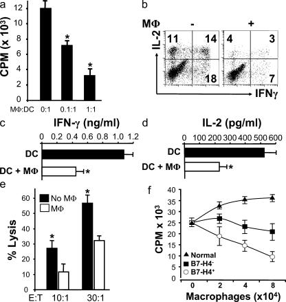
Figure 4.
Tumor macrophages suppress TAA-specific T cell immunity in vitro. (a–e) Autologous tumor ascites CD3+CD25− T cells were stimulated with Her-2/neu peptide-loaded MDC (TAA-MDC) as described in Materials and methods. (a) Tumor T cell proliferation was detected by [3H]thymidine incorporation (CPM). Results are expressed as the mean of cpm ± SEM. TAA-specific T cell IFN-γ (b and c) and IL-2 (b and d) production was detected by intracellular staining (FACS) and ELISA. (e) Tumor macrophages inhibited Her-2/neu–specific T cell cytotoxicity. MDC-activated Her-2/neu–specific T cells were effector cells, and Her-2/neu peptide-loaded T2 cells were target cells. Her-2/neu–specific cytotoxicity was determined by FACS (see Materials and methods). Tumor macrophage to TAA-MDC ratio was 1:1 for panels b–e. (f) B7-H4+ tumor macrophages significantly suppress T cell proliferation. B7-H4+ or B7-H4− tumor macrophages were sorted from malignant ascites using a FACSaria. Normal macrophages were from M-CSF–treated normal peripheral blood monocytes. Tumor ascites CD3+ T cells were stimulated with anti-CD3 antibody and blood monocytes for 3 d in the presence of different concentrations of tumor macrophages or normal macrophages. T cell proliferation was detected by [3H]thymidine incorporation. Results are expressed as the mean of cpm ± SEM. TAM, tumor macrophages; macrophage, MΦ.

