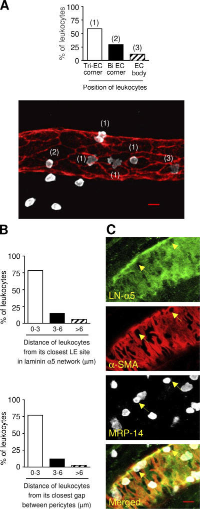
Figure 4.
Association of neutrophils in IL-1β–stimulated tissues with endothelial cell junctions, LE sites in the laminin 10 network, and gaps between pericytes. (A) IL-1β–stimulated mouse cremasteric tissues (50 ng/mouse injected intrascrotally, 4-h time point) were immunostained for the endothelial cell (EC) marker CD31 (red) and neutrophil marker MRP-14 (white). Three-dimensional images of semi-vessels of interest were captured and association of neutrophils with different EC junctions (i.e., found at tricellular or bicellular junctions) or the EC body was quantified. The graph shows the percentage of neutrophils at different positions relative to EC junctions. A representative fluorescence micrograph is shown in which examples of positions of neutrophils at tri-EC junctions, bi-EC junctions, and EC body are indicated by “(1),” “(2),” and “(3),” respectively. The corresponding bars on the graph are labeled with the same numbering scheme. For these studies, a total of 108 the percentage of neutrophils within different distance ranges from their closest LN-α5 LE sites (top) and gaps between pericytes (bottom). (C) Images show representative fluorescent micrographs of venules (semi-vessels) in IL-1β–stimulated tissues triple stained for laminin 10 (LN-α5 chain), α-SMA, and MRP-14. Selected regions of laminin LE sites are shown by arrows. Results are from four to five vessel segments/tissue (n = 6 cremaster muscles). Bar, 10 μm.

