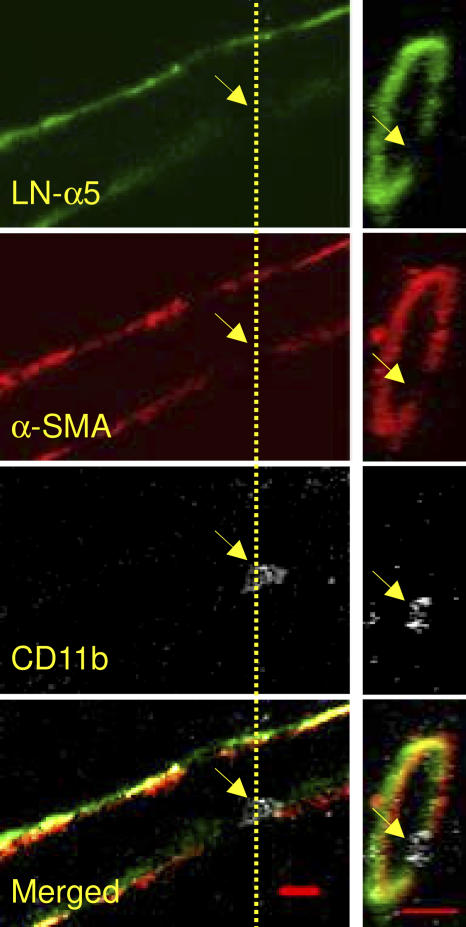
Figure 5.
In IL-1β–stimulated tissues, neutrophils migrate through laminin 10 LE sites and gaps between pericytes. IL-1β–stimulated mouse cremasteric tissues (50 ng/mouse injected intrascrotally, 4-h reaction) were immunostained for laminin 10 (LN-α5 chain), α-SMA (pericyte marker), and CD11b (leukocyte marker) and analyzed by confocal microscopy. The images on the left are from a midline optical section of a three-dimensional image of a whole venule along its longitudinal axis. The images on the right were obtained by cutting a cross section of the venule along the indicated yellow line. These representative images clearly indicate colocalization of a laminin 10 LE site, a pericyte gap, and a transmigrating leukocyte (all indicated by yellow arrows). Bar, 10 μm.

