Figure 8.
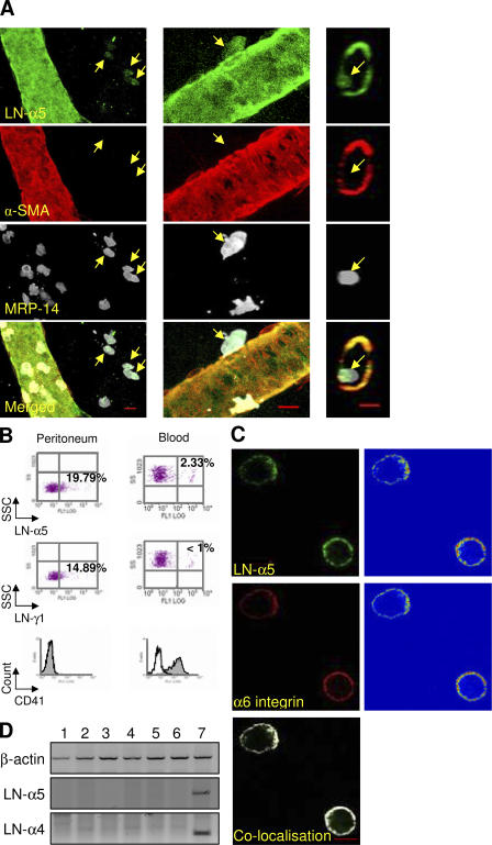
Transmigrated neutrophils carry laminin on their cell surface. (A) IL-1β–stimulated mouse cremasteric muscles were immunostained for LN-α5 chain, α-SMA (pericyte marker), and MRP-14 (neutrophil marker), and three-dimensional images of whole vessels were collected by confocal microscopy. The fluorescence micrographs show cross-sectional images and three-dimensional images of the same venular segment in which a transmigrating neutrophil (arrows) is shown to be LN-α5 chain positive. Bar, 10 μm. (B) Mice were injected i.p. with thioglycollate and peritoneal cells were harvested at 2 h after administration of the stimulus. Cell surface expression of LN-α5 and -γ1 subunits on blood or peritoneal neutrophils were quantified by flow cytometry and results are shown as scatter profiles. The numbers in each scatter profile shown in bold represent percent of positive cells. The cell samples were also stained for CD41 (platelet marker) at 4 h after stimulation. The results are representative of three independent experiments. (C) IL-1β–elicited transmigrated peritoneal leukocytes were immunostained for LN-α5 chain and α6 integrin and analyzed by confocal microscopy. Representative images of midlevel sections of cells are shown together with their corresponding intensity profiles (right). Colocalization of LN-α5 and α6 integrin was better indicated using the LSM 5 Pascal software that allows colocalized pixels of the two fluorescence channels to be visually shown by a white mask superimposed onto the original RGB image. (D) RT-PCR analysis for β-actin, LN-α5, and α4 chains from purified neutrophils (lanes 1, 3, 5) and residual mononuclear cells (lanes 2, 4, 6) harvested from thioglycollate (lanes 1–4) or IL-1β (lanes 5–6) elicited peritonitis studies. Confluent murine cardiac endothelial cells (lane 7) were used as a positive control.

