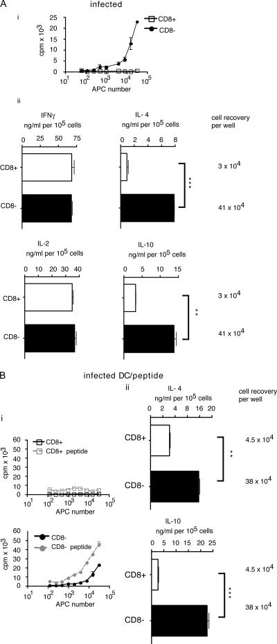
Figure 3.
CD8− but not CD8+ DCs from infected mice induced proliferation and high levels of IL-4 and IL-10 production in specific Tg CD4 T cells. (A) CD11c+CD8+ (open squares) and CD8− (closed circles) DCs from mice 7 d after infection with P. chabaudi (i) were cultured in the presence of 105 Tg T cells for 3 d, and the proliferative response was determined. CD8+ (white bars) and CD8− (black bars) splenic DCs from mice 7 d after inoculation with P. chabaudi (ii) were cultured in the presence of enriched CD4 B5 T cells. Graphs shown are the ELISAs of the mean values of a representative experiment (of three performed) with the SEM (error bars) of triplicate samples. Asterisks denote significant differences. (B) CD8+ DCs from infected mice cocultured with peptide induce significantly lower levels of IL-4 and IL-10 and fail to induce proliferation in Tg T cells. Sorted DCs from infected mice were cultured in the presence of 1 μM B5 peptide and B5 T cells and tested for Ag-specific proliferation (i) and cytokine production (ii) as in A.

