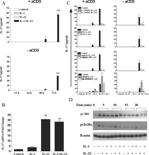
Figure 4.
IL-1 and IL-23 promote IL-17 production in the absence of TCR stimulation via stimulation of PI3K, NF-κB, and PKCθ. (A) Splenic CD3+ cells (5 × 10⁵ cells/ml) from C57BL/6 mice were incubated with 10 ng/ml IL-1β, 10 ng/ml IL-23, IL-1 and IL-23, or medium only in the presence or absence of anti-CD3, and supernatants were collected at 12, 24, 48, or 72 h for analysis by ELISA. (B) Splenic CD3+ cells from C57BL/6 mice were incubated with 1 μg/ml anti-CD3 for 2 h before stimulation with IL-1, IL-23, IL-1 and IL-23, or medium. IL-17 mRNA expression was evaluated by real-time PCR 48 h after stimulation, and the data represent the fold change for IL-17 mRNA in relation to 18S rRNA. IL-23 plus IL-1 versus IL-23 only: *, P < 0.05; **, P < 0.01; ***, P < 0.001. (C) CD3+ cells (5 × 10⁵ cells/ml) were stimulated with medium (control) or with IL-23, IL-1, or IL-23 and IL-1 with or without anti-CD3 in the presence or absence of (30-min pretreatment) LY294002 (PI3K inhibitor at 20 and 40 μM), BAY 11-7082 (NF-κB inhibitor at 4 and 8 μM), rottlerin (novel PKC inhibitor at 10 and 100 μM), UO126 (ERK inhibitor at 5 μM), or SB203580 (p38 inhibitor at 5 μM). Supernatants were collected 3 d later for analysis of IL-17 production by ELISA. IL-23 plus IL-1 versus IL-23 plus IL-1 plus inhibitor: *, P < 0.05; **, P < 0.01; ***, P < 0.001. (D) CD3+ cells were incubated with medium only or with IL-1, IL-23, or IL-1 and IL-23. Cell lysates were analyzed by Western blotting with antibodies specific for phosphorylated Akt and IκB-α or β-actin as a loading control.

