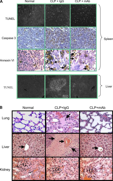
Figure 4.
Anti-HMGB1 mAb reduces serum levels of proinflammatory cytokines and organ damage without altering apoptosis in the spleen of septic mice. BALB/c mice were subjected to CLP surgery and received anti-HMGB1 mAb or nonimmune IgG administered intraperitoneally at 10 μg/mouse 24 h after CLP. Mice were killed 40 h after CLP surgery. In some experiments, between 30 and 72 h after CLP, any mice that looked very ill were killed (another mouse was killed from the control group at the same time). (A) Tissue sections of spleens (top) and livers (bottom) of normal or septic mice were prepared by using the standard formalin-fixed, paraffin-embedded procedure and were mounted on glass slides, and TUNEL assay, caspase 3 (top), and Annexin VI (top) stainings were performed. Arrows indicate the positive-staining cells. (B) Tissues were stained with hematoxylin and eosin. (top left) Normal lung shows thin alveolar septal wall and normal cellularity. (top middle) Lung from a septic mouse showing alveolar septal wall thickening, increase in cellular infiltrates, alveolar congestion, hemorrhage, and edema (arrow). (top right) Lung from a septic mouse treated with anti-HMGB1 mAb showing near normal alveoli with thin septum. (middle left) Normal liver showing a central vein (arrow) surrounded by hepatocytes and sinusoids. (middle) Liver from a septic mouse showing a congested central vein (arrow) and necrotic lesions (arrows) as revealed by the loss of cells and the structure of hepatic acinus. (middle right) Liver from a septic mouse treated with anti-HMGB1 mAb showing a central vein (arrow) and surrounding near normal hepatocytes. (bottom left) Normal kidney showing cortex with the glomerulus (G). (bottom middle) Kidney from a septic mouse showing the cortex with swelling tubule epithelia and congestion. (bottom right) Kidney from a septic mouse treated with anti-HMGB1 mAb showing swelling tubule epithelia and congestion. Data are representative of four to eight animals per group.

