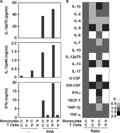
Figure 6.
IL-12 and IFN-γ production by cocultured monocytes and T cells from the patients studied and healthy controls. (A) IL-12p70, IL-12p40, and IFN-γ production, measured using classical sandwich ELISA, in a mixture of purified monocytes and T cells, as indicated upon stimulation with PHA. The results shown are representative of three independent experiments for P2 and one for P3. (B) The same coculture supernatants were analyzed for a multiplex of 16 cytokines, using the Bioplex array. Each column represents the data for one monocyte–T cell coculture system, and all four columns correspond to the same experiment. Each row corresponds to one cytokine. The gray-scale bar indicates the magnitude of cytokine expression, using the control/control (C/C) coculture system as a reference. For each data point, the amount of cytokine produced in the unstimulated system was subtracted from that produced in the PHA-activated system, and the result obtained was compared with the reference value (C/C). The production of *MCP-1 and *MIP-1β by monocytes was PHA-dependent but T cell–independent, as monocytes responded to PHA by producing large amounts of these cytokines, whereas the addition of T cells did not increase cytokine production. The defects in the production of IL-6, IL-12p70, G-CSF, IFN-γ, and MCP-1 were confirmed in three independent experiments on blood cells from P2 and one experiment on blood from P3.

