
Figure 3.
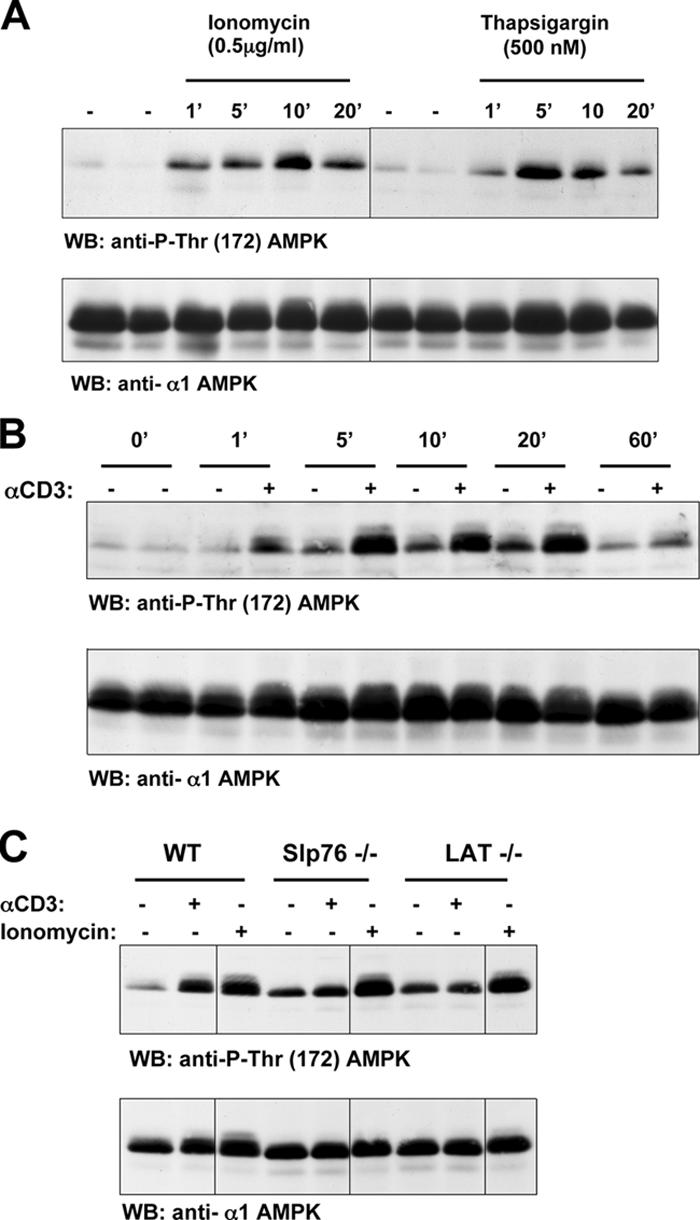
Ca2+ and TCR activation of AMPK in Jurkat cells. (A) Ionomycin and thapsigargin induce AMPK Thr-172 phosphorylation in Jurkat cells. Jurkat cells were unstimulated or treated with 0.5 μg/ml ionomycin or 500 nM thapsigargin for the indicated time periods (given in minutes). (B) TCR stimulation of AMPK Thr-172 phosphorylation in Jurkat cells. Jurkat T cells were unstimulated or treated with 10 μg/ml of the CD3 antibody UCHT1 to cross-link the TCR for the indicated time periods (given in minutes). (C) TCR stimulation of AMPK Thr-172 phosphorylation is dependent on LAT and SLP76. Wild-type, Slp76-, or LAT-negative Jurkat cells were unstimulated or treated with 10 μg/ml of the CD3 antibody UCHT1 or 0.5 μg/ml ionomycin for 5 min. (A–C) The data show Western blot (WB) analyses of cell lysates prepared from these T cells with pThr-172–AMPK or AMPKα1 antisera. Black lines indicate that intervening lanes have been spliced out.