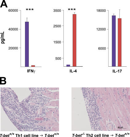
Figure 4.
Th1 as well as Th2 CD4+ T cell lines trigger autoimmune myocarditis. (A) MyHC-α–specific lines were generated by subjecting CD4+ splenocytes from immunized T-bet +/+ (blue) or T-bet −/− (red) mice to multiple rounds of antigen restimulation followed by rest in minimal IL-2. Supernatant was collected for ELISA 48 h after the third antigen restimulation. ***, P < 0.0001. One representative experiment is shown. (B) Both MyHC-α–specific Th1 cell lines (left) and Th2 cell lines (right) are pathogenic in wild-type recipient mice. Hematoxylin and eosin–stained sections: bar: 50 μm.

