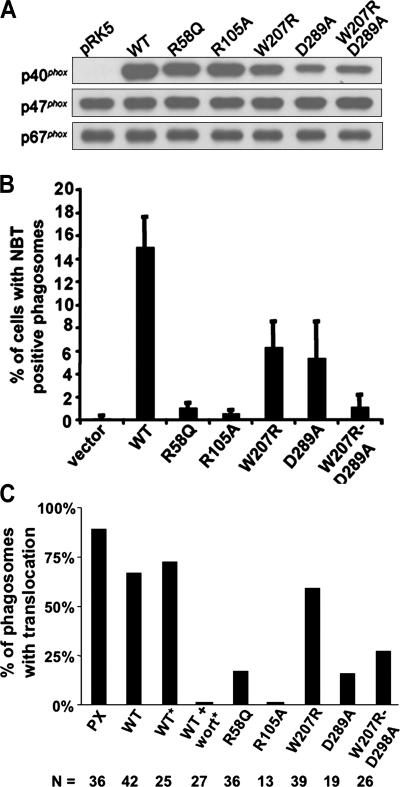
Figure 5.
Expression of p40phox mutants in COSphoxFcγR cells and effect on IgG–sheep RBC–elicited NADPH oxidase activity. Data shown is representative of at least three independent experiments. (A) Immunoblot of cell lysates from COSphoxFcγR cells transfected with 0.67 μg of either empty pRK5 or pRK5 containing cDNAs for either wild-type or mutant p40phox. Blots were probed with antibodies for p40phox, p47phox, and p67phox. (B) COSphoxFcγR cells were transfected as in A and incubated with IgG-RBCs in the presence of NBT for 30 min at 37°C. The percentage of cells with NBT+ phagosomes is shown as the mean ± SD (n = 4 except for W207R/D289A, where n = 3). (C) COSphoxFcγR cells were transfected as in A for expression of YFP-tagged wild-type or mutant derivatives of p40phox as indicated or a YFP-tagged PX domain of p40phox and incubated with IgG–sheep RBCs or with IgG latex beads (*) without or with 50 nM wortmannin, followed by confocal microscopy. Individual phagosomes were scored for either the presence (black bars) or absence of YFP-p40phox or YFP-p40PX translocation. The number of phagosomes scored for each construct is also shown. Data was collected from two to four independent experiments.

