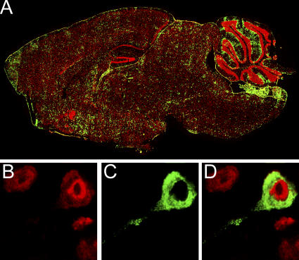
Figure 1.
Localization of LCMV in the brain of a carrier mouse persistently infected from birth. (A) Six-μm sagittal brain sections from C57BL/6 LCMV carrier mice (n = 3) at >8 wk of age were stained with a polyclonal anti-LCMV antibody (green) and a nuclear dye (red). Brain reconstructions were performed to illustrate the anatomical distribution of LCMV. Note the equal distribution of LCMV throughout the brain parenchyma. (B) High resolution analyses of LCMV-infected cells (green; C and D) in the brain parenchyma confirmed that >99% of the cells colocalized with neuronal staining (red; B and D). Overlapping fluorescence appears in D.

