Figure 2.
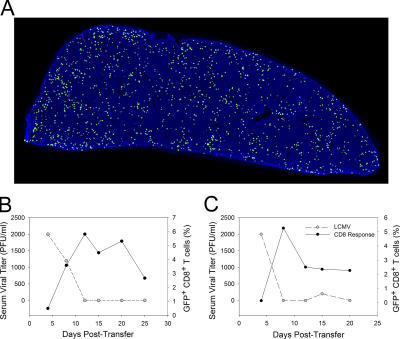
Kinetics of DbGP33–41-specific T cell expansion and viral clearance in the blood after immunocytotherapy. C57BL/6 mice seeded with 104 GFP+ (or Thy1.1+) DbGP33–41-specific CD8+ T cells and i.p. infected 1 d later with LCMV served as memory donors for immunocytotherapy experiments. (A) The distribution of GFP+DbGP33–41-specific memory CD8+ T cells (green) on a splenic reconstruction is shown for a donor mouse 45 d after infection. The illustration is a representative example (n = 3) of an entire splenic cross section. Note the even distribution of GFP+ memory cells throughout the spleen. Cell nuclei are shown in blue for anatomical purposes. (B and C) 107 memory splenocytes containing GFP+DbGP33–41-specific memory CD8+ T cells were injected i.p. into LCMV carrier mice. The viral clearance kinetics in relation to the expansion of GFP+DbGP33–41-specific T cells in the blood is shown for two representative immunocytotherapy recipients (n = 3 mice). Note that a rapid decline in serum viral titers (gray dots) coincides perfectly with the expansion of GFP+DbGP33–41-specific T cells (black dots). DbGP33–41-specific T cells are represented as the percentage of CD8+ T cells that are GFP+.

