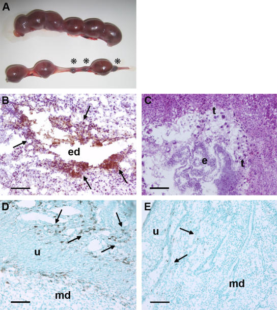
Figure 1.
Resorption of embryos, deposition of complement C3, and infiltration of monocytes in decidual tissue of DBA/2-mated CBA mice. (A) Representative uteri from mice killed at day 15 of pregnancy are shown. The top panel, from a BALB/c-mated CBA/J mouse contains larger amnion sacs and no resorptions. The bottom panel, from a DBA/2-mated CBA/J mouse, shows six amnion sacs of varying sizes and three resorptions (asterisks). (B–E) Pregnant mice were killed on day 8, and sections were stained with anti–mouse C3 (B and C) or anti–mouse F4/80 (D and E) to detect monocytes. In CBA/J × DBA/2 mice, there was extensive C3 deposition (brown, arrows; B) and monocyte infiltration (arrows) in the deciduas (D). In contrast, the decidual tissue from CBA/J × BALB/c mice showed minimal staining for C3 (C) and monocyte infiltration (E). e, embryo; ed, embryonic debris; md, maternal deciduas; t, trophoblasts; u, uterine wall. Bars, 0.1 mm.

