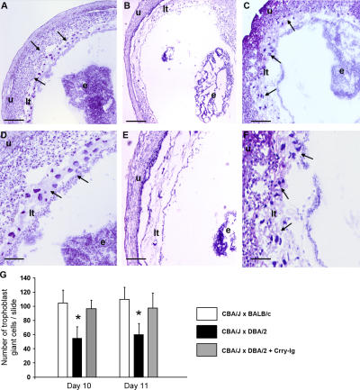
Figure 4.
Decreased trophoblast giant cells in placentas from CBA/J × DBA/2 matings. (A–F) Histologic analysis of sections of uteri from CBA/J × BALB/c matings (A and D), CBA/J × DBA/2 (B and E) matings, and (C and F) CBA/J × DBA/2 matings treated with Crry-Ig killed at day 10 of pregnancy. Trophoblast giant cells (arrows) were reduced in midtrimester placentas from DBA/2-mated CBA/J pregnancies (B and E) compared with CBA/2 × BALB/c pregnancies, and this was prevented by treatment with Crry-Ig. Sections were stained with hematoxylin and eosin. e, embryo; lt, labyrinthine trophoblast u, uterine wall. Scale bars, 0.1 mm (A–C); 0.025 mm (D–F). (G) Number of trophoblast giant cells in placentas from days 10 and 11 of pregnancy was counted by light microscopy. Data are expressed as the mean of four sections counted by two readers blinded to experimental conditions. *, P < 0.01.

