Figure 4.
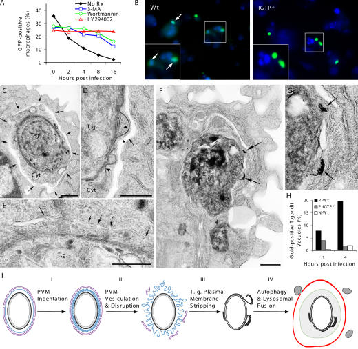
Autophagic elimination of T. gondii in primed macrophages. (A) PI3K inhibitors (100 nM wortmannin, 100 μM LY294002, and 10 mM 3-MA 30 min before and after GFP-PTG infection) attenuated rapid parasite elimination in primed WT macrophages. (B) Colocalization of GFP-PTG (green) with MDC (blue) staining in primed WT macrophages at 2 h after infection. (C and D) Autophagosome formation in primed WT macrophages. Arrows point to the autophagosome outer membrane. Note the space between the outer and inner membranes and the denuded T. gondii with part of host cytoplasm (Cyt) within autophagosomes. (D) Autophagosome formation around a partially denuded parasite. Note detachment and folding of the electron-dense plasmalemma (arrow heads). (E) Fusion of flattened vesicles (arrows) giving rise to double membrane autophagosomes. (F) Fusion of lysosomes with T. gondii–containing autophagic vacuoles. Arrows point to lysosomes containing 6-nm gold particles. (G) High magnification of the fusion event shown in F. (H) EM quantification of lysosomal fusion with T. gondii. (I) Proposed phases of T. gondii elimination in primed macrophages. Black, T. gondii plasmalemma; gray, T. gondii inner membrane complex; blue, PVM; magenta, ER; green, autophagosome inner membrane; red, autophagosome outer membrane; gray vesicles, lysosomes; bars, 500 nm.

