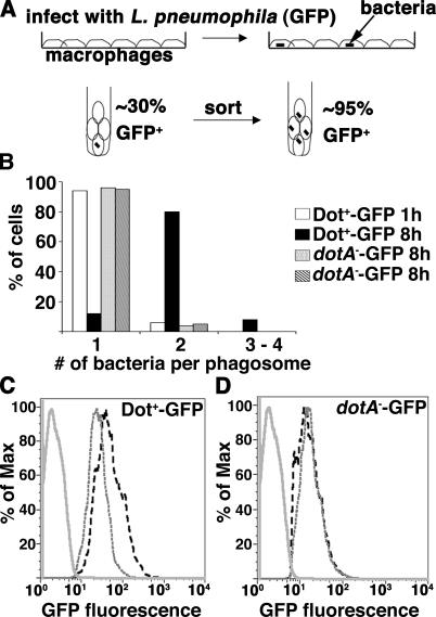
Figure 1.
Use of flow cytometry to isolate U937 cells associated with L. pneumophila. L. pneumophila strains Dot+-GFP and dotA −-GFP were introduced onto U937 macrophage monolayers at MOI = 1, allowed to incubate for 1 or 8 h, and infected cells were isolated by fluorescence sorting (Materials and methods). (A) Schematic diagram of sorting experiment. (B) One bacterial division occurs during the 8-h incubation. For each sorted GFP+ fraction, the cells were plated on coverslips, and the number of bacteria per phagosome was determined by fluorescence microscopy. (C and D) Examples of the fluorescence profile of sorted, uninfected macrophages (solid gray line); sorted, infected macrophages at 1 hai (gray dashed line); or sorted, infected macrophages at 8 hai (black dashed line) for Dot+-GFP (C) or dotA −-GFP (D), respectively. The percentage of max (% of max) indicates the number of cells relative to the peak fraction of cells.

