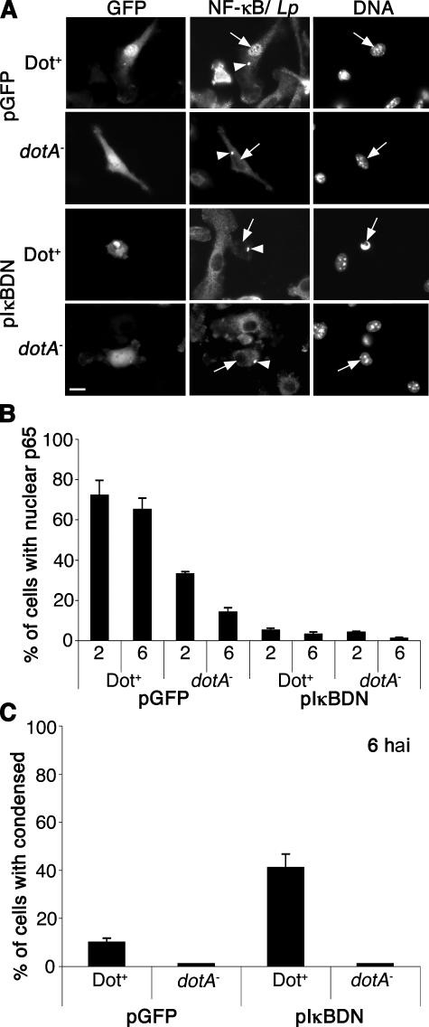
Figure 5.
NF-κB activation is required for host cell survival after L. pneumophila infection. A/J BM macrophages were transduced with retroviruses expressing either GFP or the dominant negative IκBDN-IRES-GFP construct (reference 38). Transduced A/J macrophages were then incubated with the Dot+ or dotA − strains at MOI = 1, and NF-κB p65 translocation and bacteria were visualized by immunofluorescence microscopy at 2 and 6 hai. (A) Images are examples of transduced macrophages harboring bacteria at 6 hai. NF-κB p65 and L. pnenumophila (Lp) were stained with same flour. Bacteria are marked with arrowheads, and cell nuclei are marked with arrows. Nuclear morphology was observed by Hoechst DNA staining. Bar, 10 μm. (B) NF-κB p65 translocation and (C) cell death (condensed nuclei) were observed by immunofluorescence microscopy and quantitated. Means + SE from three independent experiments are explained. A total of 100 cells harboring bacteria were counted per experiment.

