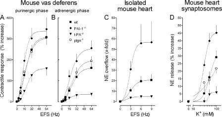
Figure 4.
Vas deferens contraction and NE exocytosis in the heart are both attenuated in t-PA–null mice, potentiated in mice with PAI-1 gene deletion, and unaffected in plasminogen-deficient mice. (A and B) Frequency response curves for the contractile responses of the isolated mouse vas deferens to electrical field stimulation (EFS; 0–64 Hz, supramaximal voltage; every 1 ms for 15 s). Peak response amplitudes of both purinergic and adrenergic phases (means ± SE [error bars]; n = 6–9) are expressed as percentages of the response to 80 mM K+. Vasa deferentia isolated from t-PA−/− mice developed markedly less tension in response to EFS than vasa from WT control mice. In contrast, vasa from PAI-1−/− mice developed more tension than vasa from WT mice. Vasa from plasminogen−/− mice developed the same tension as vasa from WT mice. (C) Coronary NE overflow from isolated mouse hearts in response to EFS (0–9 Hz; 5 V for a duration of 60 s, with pulses of 2 ms each). Hearts were perfused with buffer containing 0.1 μM desipramine, 0.1 μM rauwolscine, 1 μM atropine, and 10 μM hydrocortisone. NE overflow was significantly smaller in hearts from t-PA−/− mice than in hearts from WT mice, whereas it was significantly greater in PAI-1−/− hearts. Points are means (± SE; n = 6–9) of x-fold increases in NE overflow above basal levels. (D) K+-induced NE exocytosis in mouse heart synaptosomes. Points are means (± SE; n = 12–16) of increases in NE release above basal levels. Synaptosomes isolated from hearts of t-PA−/− mice released significantly less NE in response to K+ than synaptosomes from WT hearts. In contrast, synaptosomes from PAI-1−/− hearts released greater amounts of NE than synaptosomes from WT hearts, whereas NE exocytosis in synaptosomes from plasminogen−/− mice was not different from that of synaptosomes from WT hearts.

