Figure 3.
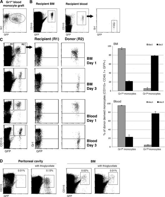
Gr1high blood and BM monocytes shuttle between blood and BM. (A) Flow cytometry analysis of a blood monocyte graft isolated from Rag−/− CX3CR1gfp mice. The GFP− cells are Ly6G+ CX3CR1− granulocytes (reference 18). (B) Analysis of recipient BM and blood 4 d after adoptive cell transfer of Gr1high blood monocytes (105 cells). Note the presence of graft-derived (GFP+) Gr1low monocytes in the blood. Fractions of graft-derived GFP+ cells of total CD115+ cells in recipient BM (0.12%) and in recipient blood (0.76%). (C) Analysis of BM and blood of WT recipients of Gr1high BM monocytes (5 × 10⁵ cells; purity: 96%) days 1 and 3 after i.v. transfer. Note the loss of Ly6C/G expression on grafted Gr1high BM monocytes. Bar diagrams summarize data obtained from three mice per time point. Note the difference of GFP intensity of BM and blood monocytes on day 1 (mean fluorescence intensity: 274.75 ± 13.9 vs. 376.6 ± 20.5; P = 0.003). (D) Flow cytometry analysis of BM and peritoneal cavity (PC) lavage of WT recipients of MACS-purified CD115+ BM monocytes (106 cells; purity: 88%) that were left untreated or had been inoculated with thioglycollate. Note the recruitment of grafted Gr1high BM monocytes to the inflamed peritoneal cavity. The data are representative of at least two independent experiments per time point.

