Figure 1.
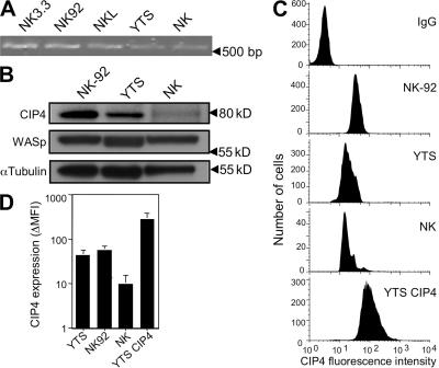
CIP4 expression in NK cells. (A) RT-PCR for CIP4 message in NK cell lines and ex vivo NK cells. (B) Western blot (10 μg of protein per lane) for CIP4 in NK92, YTS, and ex vivo NK cells, as well as WASp and α-tubulin after stripping and reprobing membranes. (C) Intracellular CIP4 FACS using CIP4 mAb or IgG clone MOPC21 (in YTS cells as a specificity control, which was comparable with IgG control for the other cell types). Ex vivo NK cells were identified by FACS in total PBMCs by costaining for CD3 and CD56 and gating on CD3−, CD56+ lymphocytes (NK). (D) The increase in CIP4 mean fluorescence intensity (MFI) over control IgG detected by FACS for YTS, NK92, ex vivo NK, and CIP4 YTS cells in three experiments and with six different donors of ex vivo cells is shown. IgG MFI was determined in parallel with each repeated assessment of CIP4. Error bars represent the SD.

