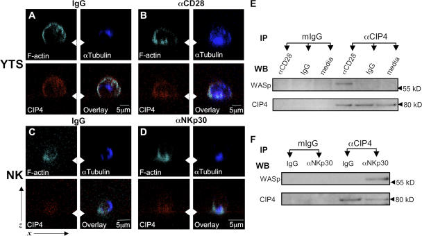
Figure 7.
Alteration in association of endogenous CIP4 after NK cell activation. YTS cells were incubated on slides coated with IgG (A) or anti-CD28 (B) and ex vivo NK cells on slides coated with IgG (C) or anti-NKp30 (D) for 30 min and evaluated via confocal microscopy in the x-z plane. The arrowheads show the plane of the slide. Fluorescence demonstrating F-actin (cyan), α-tubulin (blue), and CIP4 (red) is shown. (E) 106 YTS cells were incubated in wells containing IgG, anti-CD28 mAb, or media for 30 min and then lysed. CIP4 was immunoprecipitated using mAb anti-CIP4 and probed for WASp and CIP4 by Western blotting. Immunoprecipitation with nonspecific mIgG was performed as a control. (F) 107 ex vivo NK cells were lysed after a 30-min incubation in wells coated with IgG or anti-NKp30 mAb, and CIP4 was immunoprecipitated using mAb anti-CIP4. WASp and CIP4 were detected in immunoprecipitates by Western blotting. Immunoprecipitation with nonspecific mIgG was performed in parallel as a control. Blots represent at least three independent results.

