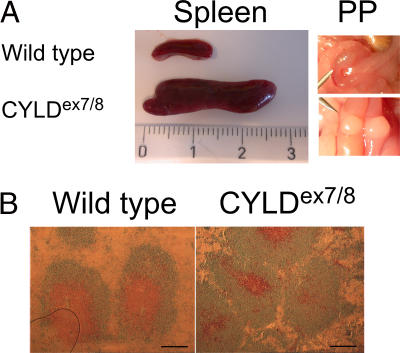
Figure 2.
Enlarged spleen and Peyer's patches (PP) in CYLDex7/8 mice. (A) Spleens and PP dissected from WT and CYLDex7/8 mice were compared. Ruler indicates the size of the organs (centimeters). (B) Cryostat sections from WT and CYLDex7/8 spleens were immunostained for B and T cell follicles with anti-B220 (blue) and anti-CD3ε (brown). Bars, 500 μm.

