Figure 8.
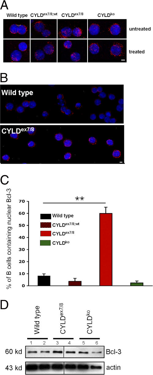
Bcl-3 accumulation in CYLDex7/8 B cells. (A) Cytospins were prepared from B cells isolated from CYLDex7/8, CYLDex7/8;wt, CYLDko, and WT mice and immunostained with anti–Bcl-3 and counterstained with Hoechst 33258. Shown are unstimulated B cells (top) and B cells treated with anti-BCR (bottom). Bar, 5 μm. (B) Same as in A, but showing more cells. Bar, 5 μm. (C) Quantification of nuclear Bcl-3 from unstimulated B cell cytospins from the indicated genotypes. For each individual genotype, 400 cells were counted and quantified for nuclear Bcl-3 localization. The frequency of nuclear Bcl-3 localization in CYLDex7/8 B cells were statistically significant (P < 0.001) compared with B cells isolated from WT, CYLDex7/8;wt, and CYLDko mice. (D) Lysates of MACS-purified B cells from the indicated genotypes subjected to Western blot analysis using Bcl-3 antibody. Duplicates for each mouse strain are shown. Actin was used as a loading control.

