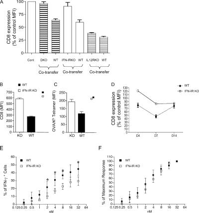
Figure 5.
Down-regulation of CD8 expression in response to LM-OVA infection is IFN-I dependent. Purified naive CD8 T cells from OT-I.PL and the indicated cytokine receptor KO OT-I strains were adoptively transferred into CD45.1 congenic B6 mice, and the host animals were infected with LM-OVA. 7 d later, recipient spleens were harvested, and WT and receptor KO OT-I cells (distinguished by staining for CD45.2 and Thy 1.1) were analyzed. (A) CD8 expression on cotransferred WT and receptor KO OT-I cells. The MFI of each group was expressed as the percentage of the MFI of CD8 staining of OT-I cells from uninfected mice. In a different experiment, CD8 expression (B) and Kb/OVA tetramer staining (C) were compared for cotransferred WT and IFN-IR−/− OT-I cells at day 7 after LM-OVA infection. Individual square symbols show CD8 expression and tetramer binding on WT and IFN-IR−/− OT-I cells in uninfected controls. (D) CD8 expression on WT and IFN-IR−/− OT1 cells as a function of time after LM-OVA infection. Dashed line represents WT cells, solid line represents IFN-IR−/− cells. (E and F) Cotransferred WT and IFN-IR−/− OT-I cells were analyzed for antigen sensitivity at day 7 after LM-OVA infection. Cells were stimulated as in Fig. 4, and the percentage of IFN-γ–positive OT-I cells (E) or the percentage of maximum IFN-γ responsiveness (F) was determined. The graphs show average responses (and SD) for three mice per group, and similar data were observed in three experiments. Stars indicate significant differences (P < 0.05).

