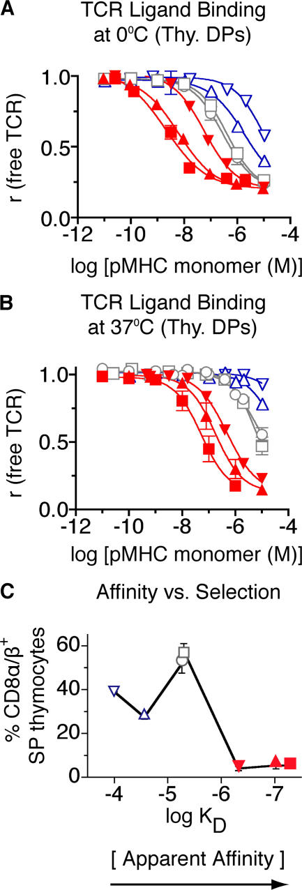
Figure 2.
Binding of soluble, monomeric pMHC ligands to live thymocytes from T1 TCR transgenic mice. Cells were incubated with several concentrations of pMHC monomers (4P-H-2Kd [▴], 4L-H-2Kd [▪], 4V-H-2Kd [▾], 4A-H-2Kd [□], 4S-H-2Kd [○], 4N-H-2Kd [▵], and 4H-H-2Kd [▿]) at 0°C (A) or 37°C (B), followed by photochemical cross-linking; the fraction of remaining free TCR was determined in a second step (two-step photoaffinity labeling method; see Material and methods and Fig. S1). (C) Selection properties of peptide variants as a function of apparent affinity [K d]. Percentages of positively selected T1 thymocytes induced by each ligand in FTOC are plotted versus their apparent affinity (Table II) for the T1 TCR. Red, negative-selecting ligands; gray, threshold ligands; blue, positive-selecting ligands. Fig. S1 is available at http://www.jem.org/cgi/content/full/jem.20070254/DC1.

