Figure 1.
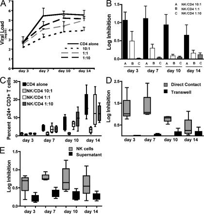
NK cells are able to mediate inhibition of HIV-1 replication in a contact-dependent manner. (A) The line graph depicts the mean change in the viral load (log10 p24 Gag in supernatant) observed in a single subject from three separate experiments in wells containing CD4+ T cells alone (continuous black line), and NK cells and autologous CD4+ T cells at a ratio of 10:1 (short dashed black line), 1:1 (long dashed gray line), and 1:10 (long dashed black line). (B) The figure represents a log reduction in p24 Gag production at different NK cell/CD4+ T cell ratios (10:1, 1:1, and 1:10) for days 3, 7, 10, and 14 (n = 5). (C) The whisker box plots show the percentage of p24 Gag+ CD3+ T cells at days 3, 7, 10, and 14 in wells containing NK cells at different NK cell/CD4+ T cell ratios (10:1, 1:1, and 1:10; n = 6). (D) The figure represents the log inhibition of HIV-1 replication observed on days 3, 7, 10, and 14 in wells where NK cells and autologous HIV-1–infected CD4+ T cells were co-cultured together (gray) or separated in a transwell experiment (black) at an NK cell/CD4+ T cell ratio of 10:1 (n = 5). (E) The whisker box plots show the log inhibition of HIV-1 replication observed on days 3, 7, 10, and 14 in wells where NK cells and autologous HIV-1–infected CD4+ T cells were co-cultured together (gray) at an NK cell/CD4+ T cell ratio of 10:1, or where the supernatant was transferred from co-cultured cells to autologous HIV-1–infected CD4+ T cells (black; n = 5). Data represent the mean of at least three experiments and standard deviations.

