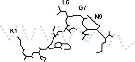
Figure 1.
Diagrammatic representation of the side chains of the HIV-1 gag peptide KRWIILGLNK bound to HLA-B27, according to the structure determined by Stewart-Jones et al. (reference 13). The α-1 helix is shown as a light blue dotted line. The floor of the peptide-binding groove below the peptide and α 2 helix are not depicted. Only the side chains of leucine 268 (L6) and asparagine 271 (N9) are exposed to the T cell receptor.

