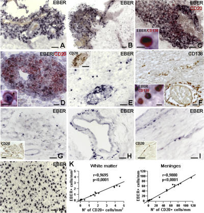
Figure 2.
Detection of EBER+ cells in the MS brain by in situ hybridization. (A–E) EBV-high MS cases. In situ hybridization for EBER shows enrichment of EBER+ cells (blue-black nuclei) in ectopic B cell follicles located in the meninges of two different MS cases (A and B). Combined in situ hybridization for EBER and immunostaining for CD20 (red surface staining) reveals a high frequency of EBER+ B cells in an ectopic B cell follicle (C) and a lower percentage of B cells expressing EBER in the large perivascular cuff of an acute white matter lesion (D; the same lesion stained for CD20 and Ki67 in an adjacent section is shown in Fig. 1 H). In the insets in C and D, an EBER+/CD138+ plasma cell and an EBER+/CD20+ B cell are shown, respectively, at higher magnification. Perivascular EBER+ (E), CD20+ B cells (inset in E), and CD138+ plasma cells (F) in a large periventricular white matter lesion (E), and some intraparenchymal infiltration by EBER+ cells (E) and plasma cells (F) in the same region. The inset in F shows CD138+ plasma cells positive for EBER inside the parenchyma. (G–I) EBV-low MS cases. EBER+ cells in the meninges (G) and around scarcely infiltrated blood vessels (H and I) in chronic active white matter lesions. The insets in G and I show CD20 immunostaining of the corresponding areas in adjacent sections. In situ hybridization for EBER in a control, EBV-associated B cell lymphoma (J). Bars: A–C, E, G, J, and insets in E, G, and I, 50 μm; D and F, 20 μm; insets in C, D, and F, 10 μm. (K) Statistically significant correlation between the number of CD20+ and EBER+ cells in the white matter (left) and meninges (right) of EBV-low (n = 12) and EBV-high (n = 8) MS cases. EBER+ and CD20+ cells were counted as described in Materials and methods.

