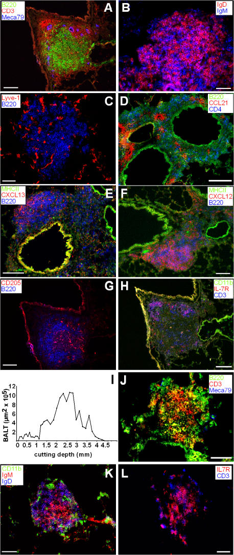
Figure 2.
Immunohistology of lungs of CCR7-deficient mice and positioning of BALT within the lung. (A–H) Cryosections of 6–8-wk-old CCR7-deficient mice were stained with antibodies as indicated. Shown are sections representative for 11 animals analyzed. (I) Lungs of CCR7-deficient mice were cut in 8-μm cryosections. Every 10th section was stained with hematoxylin and eosin. From all these sections overviews were achieved using automated image assemblies. Areas of BALT were measured. Shown is the BALT area per section in dependence of the dorsoventral cutting depth of one animal representative of six animals analyzed. (J–L) Cryosections of 6–10-wk-old gnotobiotic CCR7-deficient mice were stained with antibodies as indicated. Shown are sections representative of seven animals analyzed. Bars, 50 μm.

