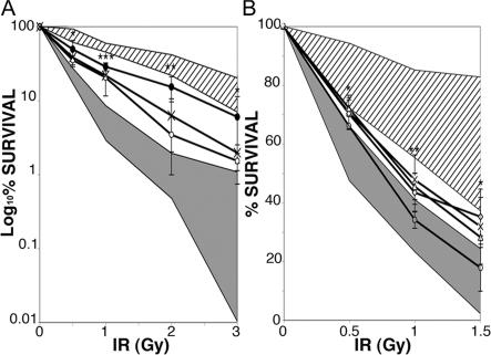
Figure 4.
Increased radiosensitivity of patients' cell lines. (A) Fibroblasts from patients (P2 [X], P3 [•], and P4 [○]) were irradiated at 0.5–3 Gy. Survival was assessed after 14 d of culture as the number of colony-forming cells compared with nonirradiated cells. Fibroblasts from two age-matched controls and fibroblasts from two Ig CSR–deficient patients with normal SHM (diagonal lines), one ARTEMIS−/−, and one A-T cell line (gray) were positive and negative controls. Results are expressed in log scale. *, P < 0.05; **, P < 0.005; ***, P < 0.001 (unpaired two-tailed Student's t test). (B) EBV B cell lines from patients (P1 [▵], P2 [X], P4 [○], and P5 [⋄]) were irradiated at 0.5–1.5 Gy. After 10 d of culture, survival was assessed as the number of positive wells (defined as viable cell colonies containing >32 cells) for each plate containing irradiated cells compared with number of positive wells for each plate containing unirradiated cells. EBV B cell lines from four controls, three AID-deficient patients, two UNG-deficient patients, and six patients with Ig CSR deficiency with normal SHM (diagonal lines), and two A-T–EBV B cell lines (gray) were used as positive and negative controls. *, P < 0.05; **, P < 0.005 (unpaired two-tailed Student's t test). Radiosensitivity of cell lines was assessed two to four times each. Results are expressed as mean ± SD.

