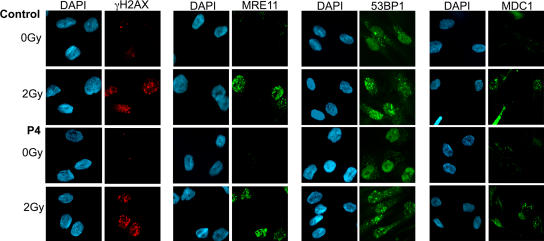
Figure 6.
Normal irradiation-induced foci formation in patients' fibroblasts. Primary fibroblasts from P4 either untreated or irradiated with 2 Gy were labeled with anti-MRE11, anti-53BP1, or anti-γH2AX mouse monoclonal antibodies and anti-MDC1 rabbit polyclonal antibody followed by anti-mouse Alexa Fluor 488, anti-mouse Alexa Fluor 546,or anti-rabbit Alexa Fluor 488. Nuclei were stained with DAPI. The same results were obtained with fibroblasts from patients P2, P3, and P4 and EBV B cell lines from patients P1, P2, and P5. Similar foci formation was observed at 5 Gy of irradiation.

