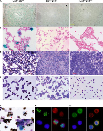
Figure 2.
Developing lymphoid cells undergo senescence. β-gal staining for thymic sections (A) and bone marrow cells (B) of Lig4−/− p53p/p, Lig4−/− p53−/−, and Lig4+/+ p53p/p mice showing positive staining only in Lig4−/− p53p/p mice. Anti-HP1γ staining of thymic sections (C) and bone marrow cells (D) of Lig4−/− p53p/p, Lig4−/− p53−/−, and Lig4+/+ p53p/p mice; positive staining was observed only in Lig4−/− p53p/p mice. (E) Bone marrow cells from Lig4−/− p53p/p mice, positive for β-gal staining, were costained with the B cell marker CD19 (dark brown cells); arrows show double-positive cells and an example of bone marrow cells from Lig4−/− p53p/p mice that are positively stained for CD19 and HP1γ (F), showing single channel of green fluorescence (HP1), red (CD19) and blue (DAPI), and merged image; (G) thymocytes staining positive for HP1γ (green), CD25 (red), and DAPI (blue). Images with wider views of both stainings are shown in Figs. S5 and S6, available at http://www.jem.org/cgi/content/full/jem.20062453/DC1.

