Figure 3.
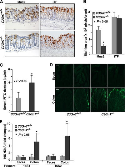
C3GnT−/− colon has reduced expression of Muc2 and impaired mucosal integrity. (A) Immunohistochemical staining of C3GnT +/+ and C3GnT −/− colonic tissues with an anti-murine Muc2 peptide antibody and with an antibody to ITF, which is a marker of fully differentiated goblet cells. Brown color indicates specific antibody binding. (B) Pixels of positive staining area per crypt were quantified using Photoshop. Six sections of three independent mice from each group were quantified. Error bars represent the mean ± the SD. (C) Serum concentration of the FITC-dextran of C3GnT +/+ and C3GnT −/− mice were measured 4 h after oral administration of FITC-dextran. Error bars represent the mean ± the SD. n = 6. (D) Representative images of fluorescent microscopic analysis of C3GnT +/+ and C3GnT −/− intestinal cryosections 4 h after oral administration of FITC-Dextran (three sections from each mouse, with four mice in each group). (E) Relative amount of 16S rDNA detected by real-time PCR using two independent sets of 16S universal primers. Data are expressed as an n-fold difference between C3GnT +/+ and C3GnT −/− mice. Error bars represent the mean ± the SD. n = 6 mice in each group. The average 16S rDNA value of C3GnT +/+ mice was expressed as 1. Bars: (A) 100 μm; (D) 50 μm.

