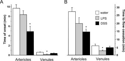
Figure 4.
DSS and LPS enhance thrombus formation in arterioles and venules, respectively. (A) Time of onset of thrombus formation in cremaster arterioles and venules exposed to light/dye. (B) Time to cessation of flow in the same vessels after light/dye exposure. Values are reported as means ± SE. *, P < 0.05 versus water.

