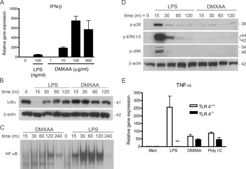
Figure 1.
DMXAA preferentially induces IRF-3–mediated gene expression. (A) Peritoneal macrophages from C57BL/6 mice were stimulated for 2 h with 100 ng/ml LPS or increasing concentrations of DMXAA, as shown. Total RNA was extracted and subjected to reverse transcription followed by quantitative real-time PCR, as described in Materials and methods. (B) Primary mouse macrophages were stimulated with medium alone, 100 ng/ml LPS, or 100 μg/ml DMXAA for the indicated times. Total protein was collected and subjected to SDS-PAGE, followed by Western blotting with anti-IκBα antibody. Protein molecular masses (in kD) appear at the right. (C) RAW 264.7 macrophages were stimulated with medium alone, 10 ng/ml LPS, or 1 mg/ml DMXAA for the indicated times. Nuclear extracts were prepared and subjected to EMSA with an NF-κB–specific labeled oligonucleotide. (D) Peritoneal macrophages from C57BL/6 mice were stimulated with medium alone, 100 ng/ml LPS, or 100 μg/ml DMXAA for the indicated times. Western blotting for activated MAPK was performed on whole-cell lysates. β-Actin was used as a loading control. Protein masses (in kD) appear at the right. (E) Peritoneal macrophages from TLR4+/+ and TLR4−/− were stimulated with medium alone, 100 ng/ml LPS, or 100 μg/ml DMXAA for 2 h. Total RNA was collected and analyzed by quantitative real-time PCR, as in A. Results represent the mean ± SE for at least three independent experiments. **, P < 0.01.

