Figure 5.
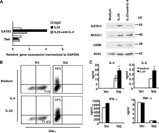
The function of IL-25 in early Th cell activation and in effector Th2 cells. (A) Naive T cells were stimulated with anti-CD3 and anti-CD28 with or without IL-25 or with the addition of anti–IL-4 antibody. Cells were collected at 72 h for real-time PCR or 48 h for Western blot analysis. (B) Purified naive T cells from transgenic OT-II mice were differentiated into Th1 and Th2 cells. After 5 d of culture, cells were washed and restimulated with anti-CD3 with or without IL-25 for 3 d to assess the levels of IL-4 and IFN-γ expression by intracellular cytokine staining. The percentages in the figure represent cells stained positive for each cytokine indicated. (C) Purified naive T cells from C57BL/6 mice were stimulated with anti-CD3 and anti-CD28 with the same treatment as in B. After 5 d of culture, Th1 and Th2 cells were washed and restimulated with anti-CD3 with or without IL-25 for 2 d to assess the levels of IL-4, IL-5, IFN-γ, and TNF-α expression by ELISA. Data are presented as mean values + SD. *, P < 0.05.

