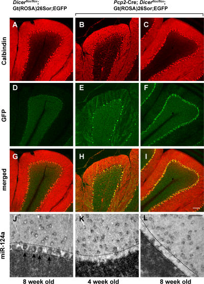
Figure 1.
Dicer inactivation in postnatal Purkinje cells. 20-μm-thick cerebellar sections of 8-wk-old control Dicerflox/flox; Gt(ROSA)26Sor;EGFP (A, D, and G), and experimental Pcp2-Cre; Dicerflox/flox; Gt(ROSA)26Sor;EGFP mice at 4 (B, E, and H) and 8 (C, F, and I) wk of age are shown. To visualize Purkinje cells, the cerebella sections were stained with anti-calbindin antibody (red). Purkinje cells expressing Cre at levels inducing loxP recombination were visualized by expression of eGFP using an anti-GFP antibody (green). The expression of brain-specific miRNA miR-124a in 8-wk-old control (J) and Pcp2-Cre; Dicerflox/flox; Gt(ROSA)26Sor;EGFP mice at 4 (K) and 8 (L) wk of age was detected by in situ hybridization. Arrows indicate individual Purkinje cells, and the Purkinje cell layer is indicated by dashed lines. Bars: A–I, 100 μm; J–L, 50 μm.

