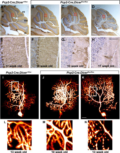
Figure 3.
Postnatal inactivation of Dicer leads to cerebellar degeneration. 12-μm-thick saggital cerebellar sections of 17-wk-old control Pcp2-Cre; Dicer+/flox mice (A and E) and 10- (B and F), 13- (C and G), and 17- (D and H) wk-old experimental Pcp2-Cre; Dicerflox/flox mice are shown. Purkinje cells were visualized by immunohistochemistry using an antibody to calbindin (brown). Sections are counterstained using Nissl stain (blue). Cerebellar lobules are indicated by roman numerals. The sections outlined by red rectangles in A–D are amplified in E–H. Morphological changes in Dicer-deficient Purkinje cells were visualized using two-photon images after intracellular injection of the calcium-sensing dye Fura 2. 13-wk-old control Pcp2-Cre; Dicer+flox (I and L) and 10- (J and M) and 13- (K and N) wk-old Pcp2-Cre; Dicerfloxflox mice are shown. The individual images were stacked and visualized using Imaris software. The sections outlined by white rectangles in I–K are shown in more detail in L–N. Bars: A–D, 1 mm; E–H, 50 μm; I–K, 20 μm; L–N, 5 μm.

